Uveitis/OID is the Third Leading Cause of Blindness

By C. Stephen Foster, MD, FACS, FACR

Did you know that Uveitis/OID is the third leading cause of blindness in developed countries? It is the third leading cause of blindness because patients are left on chronic steroid therapy, which causes vision-robbing problems of glaucoma and cataract, or they are not completely controlled by their inflammation. Chronic low grade or severe inflammation will cause macular edema, optic nerve damage, glaucoma, cataract, synechiae, and other vision-robbing complications.Dr Foster Patient

The American Uveitis Society developed guidelines for care that involve a step ladder approach to treatment moving patients from steroids. Such therapies include nonsteroidal anti-inflammatories, immunosuppressive chemotherapy, or biologics when the drug categories fail to prevent unnecessary blindness.

#uveitis #ocularinflammatorydisease #oid #glaucoma

Looking Forward to 2020: The Year of Vision a Self-Reflection

By Stephen D. Anesi, MD, FACS

This time of year is always a time for reflection, but also a time to look to the future. As we enter into this new decade, starting with the year 2020, I wanted to share some of the thoughts I have had during my 10 years spent at MERSI. First and foremost, I have had the immense pleasure of meeting and getting to know the people of New England, a place I am very proud to now call my home. And I also have been very fortunate to be called upon to help those who have traveled from distant states or countries to Boston to seek our care. Our patients, whether afflicted with the myriad of problems we are known to specialize in treating as a tertiary care center in ocular inflammatory disease, or suffering from other significant eye disease that may rob them of vision just the same, are the reason we keep doing what we do. We have been entrusted with their care, and there is no greater joy I have found in my career than each moment when a patient is helped to the point where they are able to now go about their lives without concern for their eyes or vision. And there is no greater frustration than when, despite my best efforts, a patient is still unable to experience this.

For many, the prospect of 20/20 vision is a goal they aspire to achieve. “Perfect vision.” What does this mean? It means that at 20 feet, a patient can see things that another person with “normal” or “perfect” vision can see at 20 feet. The same as 20/40 means they must come up to 20 feet to see what “normal” vision allows at 40 feet, and 20/15 means they are able to stand back at 20 feet to see what “normal” vision allows at only 15 feet. These numbers are so critically followed by many who come through our care, and small changes, which may not necessarily be medically “significant”, may make a very large difference to the person directly affected by those changes.

As physicians we are often expected to shoot for the stars, to be perfect, to fix everything – but in truth many MDs are often satisfied if the result achieved is at least up to the “standard of care”. For us physicians, I actually believe it is critical that we also remember that it is neither our needs nor expectations that are the only ones that matter, but those of the patient as well. And those needs and expectations may vary considerably… If a patient is left with near but not quite 20/20 vision, despite a very difficult course or prognosis, they may still be disheartened for not reaching that seemingly crucial endpoint. As if they somehow fell short. Yet there are others who become overjoyed to finally be able to see something recognizable at all. And then what if those needs go beyond vision?

What many do not know or come to realize is that 20/20 IS just a number… just one metric by which we as ophthalmologists judge how well an eye is able to function. The key word here is function, for vision is not the only problem we as ophthalmologists strive to manage. The overall well-being of the eye extends far beyond being 20/20, or even vision for that matter. One may reach the 20/20 level while being tested in the dark with a white background, but go out into the real world and be functionally incapacitated by some other deficit. Having 20/20 vision may not help someone see inside a darkened room after coming in from outside because of retinal disease or glaucoma, or gain back the independence of driving at night or in the rain because of overwhelming glare and haloes from cataract or astigmatism, or even still relieve debilitating ocular discomfort that prevents one from being able to concentrate or do anything at all. And these are only a few of the problems we, as doctors and patients together, face in the fight against eye disease.

Another thing many don’t come to realize is how much of an effect patients have on their doctors. I continue to learn from the people I have helped, sometimes years after I have met them. The strength, the courage, and most of all the will to continue on even when it seems there is no hope. It’s an inspiration. On both sides of the spectrum.

A patient of mine with uveitis for several decades presented to me with one remaining eye with any potential for vision, legally blind with severe astigmatism and a dense inflammatory cataract no other surgeon wanted to remove due to the risk involved. Despite the difficulty of the surgery, and the low chance it would even improve his vision due to previous retinal and optic nerve damage, he was motivated to achieve the best possible outcome, including pursuing correction of the astigmatism with a special lens. Today he regularly visits with 20/60 vision in his only seeing eye with minimal correction – and is absolutely thrilled because now, even though far from 20/20, his eye is able to function to allow him to see more, and do more, than he had been able to in many years. Needless to say, this is an outcome that would leave any doctor or patient ecstatic, as it continues to do for both he and myself each and every visit since that outcome was achieved.

Another patient of mine has had 20/20 vision in both eyes for as long as I can remember, but this was not her major concern. For despite the number, her vision was not clear, and worse, she has constant pain from scleritis and orbital inflammation. Pain to the point where life often comes to a standstill because of the inability to think or do or feel anything else, often leaving her unable to function in any capacity at all. Worse still, her case is among the most difficult to treat I can also remember. The thought of even coming to visits is at times an overwhelming burden, but a strong will and motivation to improve continues to prevail. And as she continues to battle this difficult disease, I too am motivated to continue, absolutely without end, to try to find some means of achieving relief for her so that one day she can again enjoy the life she greatly deserves.

These two brave patients help me remember some important lessons I have learned. No two patients, nor their eyes, are the same. Nor is any outcome taken for granted. Their needs are their own. What they value, crucial. What we may call routine care is a “miracle” to another. And what we consider a heroic effort may end in failure. As long as motivation remains, by both patient and doctor, to improve upon the hand dealt, I will continue moving toward that end in any capacity I am able. My goal as an ophthalmologist and ocular immunologist moving forward will remain thus… to help each patient and eye, not only just to see, but to function in a way that makes life easier and more enjoyable for the person who has entrusted me with their care. No matter what.

On behalf of myself and the other doctors and the entire staff at MERSI, I thank you for your continued faith in us to be there to help you however we can in whatever struggle you face, and I wish you all a very happy and healthy new year and decade to come!

#ophthalmology #uveitis #scleritis

Children’s Eye Health and Safety Month: Healthy Eye Vision Screening

By C. Stephen Foster, MD, FACS, FACR and Frances Foster, MS, NP

In Highlight of Children’s Eye Health and Safety Month, We Advocate, of course, eyewear protection and sunglasses, but will focus on the importance of vision screening in this blog.

Undetected and untreated vision problems can interfere with a child’s ability to learn in school and participate in sports. The earlier a vision problem is diagnosed and treated, the less it will impact an individual’s quality of life. Vision screening programs are intended to identify children who may have undetected vision problems like uveitis, especially in children with juvenile idiopathic arthritis. The recommendation for patients with JIA who are high risk for uveitis need screening with an ophthalmologist every 3 months. We highly recommend vision screening in schools, but of course, a vision screening cannot be relied upon to provide the same results as a comprehensive eye and vision examination. A pediatrician or other primary care physician may do a vision screening as part of a school physical. Vision screenings are often part of local health fairs put on by hospitals, social service agencies or fraternal groups like the Lions and Elks clubs. There are limitations to a vision screening test and cannot substitute for a comprehensive eye exam. The major limitation by American Academy of Ophthalmology is that most vision screening tests are only for distance visual acuity. While the ability to see clearly in the distance is important, it does not indicate how well the eyes focus up close or work together. Nor does it give any information about the health of the eyes. Some screenings can include a test for farsightedness and eye coordination, but even these additional screening tests can miss eye health problems.

A comprehensive eye and vision examination are always best.  An ophthalmologist can conduct a comprehensive eye and vision examination as well as make a definitive diagnosis and prescribe treatment. A comprehensive eye and vision examination include:

  • Patient and family health history
  • Visual acuity measurement with depth perception, color vision, peripheral (side) vision and the response of the pupils to light
  • Assessment of refractive status to determine the presence of nearsightedness, farsightedness or astigmatism
  • Evaluation of eye focusing, eye movement abilities
  • Additional tests as needed

Frequency of Routine Vision Screening – American Academy of Ophthalmology (AAO) recommendations:

  • New infant
  • Once from age 6-12 months
  • Once between the age of 3-5
  • School-age every 1-2 years

The recommendation from for patients with JIA who are high risk for uveitis need screening with an ophthalmologist every 3 months.

References:

Gudgel, D. (2014, August 4). Eye Screening for Children. Retrieved from https://www.aao.org/eye-health/tips-prevention/children-eye-screening

Eye Health Observances. (. (2019, August 13). Retrieved from https://www.aao.org/newsroom/observances

Recommended Eye Examination Frequency for Pediatric Patients and Adults. (2019, August 13). Retrieved from

https://www.aoa.org/patients-and-public/caring-for-your-vision/comprehensive-eye-and-vision-examination/recommended-examination-frequency-for-pediatric-patients-and-adults

#childrenseyehealth #visionscreening #ophthalmologist #uveitis #juvenileidiopathicarthritis #jia #aao #aoa

Juvenile Idiopathic Arthritis and Uveitis: What is it and what is its effect on the eye?

by C. Stephen Foster, MD, FACS, FACR

Uveitis is a serious complication of juvenile idiopathic arthritis (JIA). Approximately 6% of all cases of uveitis occur in children, and up to 80% of all cases of anterior uveitis in childhood are associated with JIA. Although remarkable progress has been made in the care of patients with JIA-associated uveitis since the development of corticosteroids for systemic and ophthalmic use in the 1950’s, up to 12% of children with uveitis associated with pauciarticular JIA still develop permanent blindness as a result of low-grade chronic intraocular inflammation. Ironically, these children are often under careful observation by ophthalmologists who may opt to tolerate low-grade ocular inflammation, hoping to avoid the development of corticosteroid-induced ocular adverse effects such as cataracts and glaucoma. The vision-robbing consequences of low-grade uveitis occur extremely slowly, typically over a period of 4 to 8 years, and the end result is clear: even low-grade uveitis may lead eventually to ocular damage, including band keratopathy, maculopathy (macular edema, macular cysts, epiretinal membrane), glaucomatous optic neuropathy, and cataract formation from chronic inflammation and corticosteroid therapy. Although surgical treatment of cataract and glaucoma is remarkably successful in the general population, it is consistently less so in patients with JIA-associated uveitis.

Further reduction in the occurrence of irreversible blindness secondary to uveitis in patients with JIA depends on early diagnosis of iridocyclitis, facilitated by mandatory vision screening programs in day care centers and schools, and on the use of therapeutic algorithms that include methotrexate and other immunomodulators to eradicate intraocular inflammation. Patients with JIA-associated uveitis should be promptly referred to colleagues experienced with such therapy before permanent ocular damage develops. Our hope is that cooperation among physicians of all specialties caring for patients with JIA will reduce the ocular morbidity and blindness secondary to JIA-associated iridocyclitis.
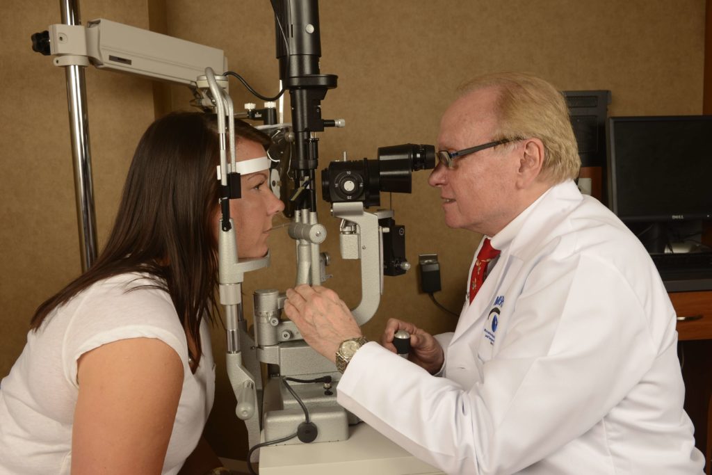
Epidemiology

The prevalence of JIA in the United States has been estimated recently at 30,000 to 50,000 cases. JIA is more common in girls, with a female-to-male ratio of 3:2. There are 3 types of JIA onset: systemic, which constitutes 11% to 20% of cases; polyarticular (5 or more joints), 17% to 40%; and pauciarticular, 40% to 72%. The peak age at onset of JIA is between 2 and 4 years. The incidence of iridocyclitis in patients with JIA ranges from 8% to 24% and varies among sub-groups of JIA. Uveitis occurs both in children with JIA who are antinuclear antibody (ANA) positive and in those who are ANA negative. Approximately 78% to 90% of patients with JIA-associated iridocyclitis have pauciarticular arthritis; 90% of these patients are ANA positive. Between 7% and 14% of JIA patients with uveitis have polyarticular arthritis; 2% to 6% have systemic arthritis. Uveitis precedes the onset of arthritis in approximately 6% of cases and may be detected at the time of initial diagnosis of arthritis. The majority of patients develop iridocyclitis within 4 to 7 years after JIA onset; the average age at the time of diagnosis of JIA-associated iridocyclitis is 6 to 8 years. The highest risk of iridocyclitis is within 2 years after the onset of arthritis and declines considerably after 8 years have elapsed from the onset of JIA. In some patients, however, the interval between onset of arthritis and uveitis may exceed 20 years. In general, little or no correlation exists between arthritis activity and the presence of uveitis. The uveitis associated with JIA is typically anterior, involving the iris and ciliary body, and generally affects both eyes.

Diagnosis and Course

The eyes of patients with JIA-associated uveitis commonly appear normal (not red or inflamed) on external examination and routine ophthalmoscopy. Because patients with JIA are young, they may not notice or report small visual changes that are slowly developing as a result of active inflammation. Therefore, current guidelines recommend that children whose age at onset of JIA is less than 7 years and who do not have known iridocyclitis should have a complete ophthalmologic examination including slit lamp evaluation, every 3 to 4 months if they have pauciarticular or polyarticular JIA and ANA positivity, every 6 months if they have pauciarticular or polyarticular JIA but ANA negativity, and every 12 months if they have systemic JIA. However, Candell, Chalom and colleagues recently reported that, in a retrospective study, although the prevalence of uveitis was lower in ANA-negative patients compared with ANA-positive patients, ocular complications were more common in children with uveitis who were ANA negative. These authors speculated that perhaps ANA-negative children were screened less intensively and thus fared worse. Patients with JIA are considered at low risk for developing iridocyclitis 7 years after the onset of their arthritis but should have yearly ophthalmologic examinations indefinitely thereafter. Once iridocyclitis is diagnosed, the managing ophthalmologist can determine the frequency of visits, depending on the severity of uveitis, its response to therapy, and the treatment used.

Therapy

There are no randomized controlled trails evaluating treatment strategies for JIA-associated uveitis. Observational studies and clinical experience have led us to advocate a treatment philosophy of tolerating no active inflammation at any time in the eyes of children with JIA-associated iridocyclitis. We use a “stepladder” algorithmic approach of progressive aggressiveness of treatment to achieve that goal. The cornerstone of initial management is topical steroid therapy (prednisolone 1%). Regional injection steroids (triamcinolone acetonide) and systemic steroids (prednisone) are often used simultaneously in the initial care of a patient with JIA-associated iridocyclitis. If the inflammation persists after 90 days of treatment and attempted withdrawal of oral and topical steroids, chronic use of an oral NSAID(nonsteroidal anti-inflammatory drug) is indicated. Naproxen (available in suspension), 10 to 15 mg/kg per day in 2 divided doses, is a common first choice; tolmetin, 20 to 30 mg/kg per day in 4 divided doses is another choice. These 2 agents have the longest safety record in childhood use. However, pediatricians and pediatric rheumatologists may choose from a wide range of other NSAIDs and dosages for their patients. We usually prescribe a histamine H2-receptor antagonist (ranitidine or famotidine) or the synthetic prostaglandin E1analog misoprostol as prophylaxis for gastritis or ulcerative symptoms for patients who receive NSAIDs, especially if an NSAID is given with a systemic steroid. The Cox-2 specific NSAIDs, such as Celebrex, may become especially appealing for long-term care.

In approximately 30% of cases, JIA-associated iridocyclitis is not satisfactorily controlled by conventional anti-inflammatory treatments, and iridocyclitis recurs whenever steroids are withdrawn despite the long-term use of an oral NSAID. We believe that at this stage advancing to low-dose, once-a-week methotrexate therapy (o.3-o.5 mg/kg per week; typically, 7.5-25 mg per week) should be considered. The dosage of methotrexate can usually be increased to a maximum of 40 mg weekly to control the iridocyclitis if the standard starting dose is not adequate. Therapy with methotrexate, a folic acid antagonist, has had an exceptional safety and efficacy record as used by rheumatologists to care for children with the joint manifestations of JIA. Published data and clinical experience indicate that immunomodulatory agents have considerably fewer adverse effects than systemic corticosteroids when used as single agents in relatively low doses for the treatment of JIA-associated uveitis. The mechanisms of action of methotrexate in JIA are not well understood. However, methotrexate seems to have both anti-inflammatory and immunosuppressive actions. The potential for drug-induced adverse effects, such as bone marrow suppression, hepatic toxicity, pneumonitis, and oral ulcerations exists. Clinical studies have shown that the concurrent administration of folic acid or folinic acid with methotrexate does not reduce the efficacy of the drug but might alleviate some of its potential toxicities. The concurrent use of folic acid, 1 mg/d, the close involvement of the physician in longitudinal care, and the appropriate monitoring of hematologic parameters and hepatic enzyme levels minimize the risk of adverse effects. We believe that such careful management accounts for the safety record of low-dose, once-weekly methotrexate. There is no known risk of sterility with low-dose methotrexate therapy. The risk of increased susceptibility to a malignancy later in life also is probably not significant in children with JIA treated with methotrexate. There have been reports of Epstein-Barr virus associated lymphoma and lymphoproliferative disorders occurring in patients with adult RA receiving low-dose methotrexate therapy; discontinuation of methotrexate therapy often leads to complete remission. The authors of these studies acknowledge that methotrexate probably has no direct oncogenic effect itself, but may promote or enhance malignancies associated with Epstein Barr Virus infection in individuals with disorders such as RA. We know of no such predisposition in JIA patients.

When methotrexate treatment fails or is not tolerated, other immunomodulators, such as azathioprine (1-2 mg/kg per day), cyclosporine (2-5 mg/kg per day), mycophenolate mofetil (mg/m2 body surface area bid), or chlorambucil (0.10-0.16 mg/kg per day) may replace methotrexate or be used adjunctively to achieve the goal of total quiescence of ocular inflammation. However, the frequency of failure of methotrexate treatment is quite small. Infliximab or Adalimumab may also be considered.

#jia #juvenileidiopathicarthritis #uveitis #iridocyclitis #pauciarticulararthritis

Cataract Surgery and Uveitis

by C. Stephen Foster, MD, FACS, FACR

Cataract develops in patients with uveitis because of the uveitis itself and also because of the steroids which is the cornerstone of treating uveitis. Cataract developing in an eye with a history of chronic or recurrent uveitis has historically been called cataracta complicata, and, indeed, the uveitic cataract is complicated cataract. It is complicated both from the standpoint of technical aspects of the surgery itself (limited access secondary to posterior synechiae, pupillary membrane, and pupillary sphincter sclerosis, iris delicacy and vascular abnormalities, and pre-existing glaucoma), and also because of the high likelihood of an exuberant postoperative inflammatory response which can ruin the desired surgical outcome.

Increasing availability of more delicate microsurgical techniques, through the use of pupil expanders, visco elastic material, small incision phacoemulsification techniques, etc. has dramatically reduced the misadventures that use to be so common. Yet, despite these advances, surgeons are frequently still disappointed with the visual outcome of cataract surgery in the patient with a history of uveitis. Poor visual outcome typically occurs as a result of two things: damage done to the macula or optic nerve long before the time for cataract surgery has arrived, through the consequences of recurrent or chronic, even “low grade” inflammation; and significant ongoing chronic or recurrent inflammation which sabotages an initially good visual result from cataract surgery. Both these problems are avoidable, but avoidance of structural damage to areas of the eye critical for good vision requires a philosophy, on the ophthalmologists’ part, of total intolerance to chronic or recurrent inflammation, achieving the goal of complete freedom of inflammation through a stepladder algorithm approach in aggressiveness of therapy.

Indeed, prevention of cataract development in the first place often derives from such a philosophy of total intolerance of any inflammation. And postoperative inflammatory damage which sabotages an initially good visual outcome occurs, generally, if the patient is prepared ahead of surgery with treatment techniques that prevent exuberant inflammation postoperatively, and prevent a recurrence of inflammation or a continued low grade chronic inflammation longitudinally following surgery.

This e-mail, including any attachments, is meant only for the intended recipient and may be a confidential communication or a communication privileged by law. If you received this e-mail in error, any review, use, dissemination, distribution, or copying of this e-mail is strictly prohibited. Please notify the sender immediately of the error by return e-mail and please delete this message from your system. Any opinions expressed in this email are those of the individual and not necessarily of Massachusetts Eye Research and Surgery Institution, PC or The Ocular Immunology and Uveitis Foundation. Thank you in advance for your cooperation.

#cataractsurgery #uveitis #steroids #uveiticcataract

Cataract Surgery and Uveitis

Cataract Surgery and Uveitis
C. Stephen Foster, M.D., F.A.C.S, F.A.C.R.

Cataract develops in patients with uveitis because of the uveitis itself and also because of the steroids which is the cornerstone of treating uveitis. Cataract developing in an eye with a history of chronic or recurrent uveitis has historically been called cataracta complicata, and, indeed, the uveitic cataract is complicated cataract. It is complicated both from the standpoint of technical aspects of the surgery itself (limited access secondary to posterior synechiae, pupillary membrane, and pupillary sphincter sclerosis, iris delicacy and vascular abnormalities, and pre-existing glaucoma), and also because of the high likelihood of an exuberant postoperative inflammatory response which can ruin the desired surgical outcome. But the increasing availability of more delicate microsurgical techniques, through the use of pupil expanders, visco elastic material, small incision phacoemulsification techniques, etc. has dramatically reduced the misadventures that use to be so common.

Yet, despite these advances surgeons are frequently still disappointed with the visual outcome of cataract surgery in the patient with a history of uveitis. This typically occurs as a result of two things: damage done to the macula or optic nerve long before the time for cataract surgery has arrived, through the consequences of recurrent or chronic, even “low grade” inflammation; and significant ongoing chronic or recurrent inflammation which sabotages an initially good visual result from cataract surgery. Both these problems are avoidable, but avoidance of structural damage to areas of the eye critical for good vision requires a philosophy, on the ophthalmologists’ part, of total intolerance to chronic or recurrent inflammation, achieving the goal of complete freedom of inflammation through a stepladder algorithm approach in aggressiveness of therapy. Indeed, prevention of cataract development in the first place often derives from such a philosophy. And postoperative inflammatory damage which sabotages an initially good visual outcome occurs, generally, if the patient is prepared ahead of surgery with treatment techniques that prevent exuberant inflammation postoperatively, and prevent a recurrence of inflammation or a continued low grade chronic inflammation longitudinally following surgery.

The exact details of surgical technique, above and beyond the routine required for adequate access to the lens and control of inflammation is very much patient and disease-specific. For example, many (indeed, perhaps even most) patients with a history of uveitis can have, as part of the surgical plan, implantation of a posterior chamber intraocular lens implant; exceptions to this generalization exist, for example, in most patients with uveitis on the basis of juvenile idiopathic arthritis, and in patients for whom recurrent inflammation episodically through time is unpredictable and generally not preventable (i.e., patients with sarcoidosis, or even with patients with a history of multiple recurrences of toxoplasma retino-choroiditis.) Also, the exact details of which glaucoma procedure to perform, in the patient who needs glaucoma surgery in conjunction with cataract surgery, is also somewhat disease-specific and patient-dependent. And, finally the issue of simultaneous pars plana vitrectomy is very much disease-dependent. We believe, for example, that most patients with JIA-associated iridocyclitis and patients with pars planitis or uveitis that has been characterized by great “vitritis” or multiple recurrences affecting the posterior segment very much benefit from primary pars plana vitrectomy at the time of the cataract surgery.

For further reading on this subject may I suggest the following references:

REFERENCES:

  1. Foster CS, Barrett F: Cataract Development and Cataract Surgery in Patients with Juvenile Rheumatoid Arthritis-associated Iridocyclitis. Ophthalmology 1993; 100:809-817.
  2. Foster CS, Fong LP, Singh G: Cataract Surgery and Intraocular Lens Implantation in Patients with Uveitis. Ophthalmology 1989; 96:281-288.
  3. Foster RE, Lauder CY, Meisler DM, et al: Extracapsular cataract extraction with posterior chamber intraocular lens implantation in uveitis patients. Ophthalmology 1992; 99:1234-1241.
  4. Gee SS, Tabbara KF: Extracapsular cataract extraction in Fuchs’ heterochromic iridocyclitis. Am J Ophthalmol 1989; 108:310-314.
  5. Kaufman AH, Foster CS: Cataract extraction in pars planitis patients. Ophthalmology1993; 100:1210-1217.
  6. Seamone CD, Deschenes J, Jackson WB: Cataract extraction in uveitis: comparison of aphakia and posterior chamber lens implantation. Can J Ophthalmol 1992; 273:1231-124.

Juvenile Idiopathic Arthritis and Uveitis: What is it and what is its effect on the eye?

Juvenile Idiopathic Arthritis and Uveitis
C. Stephen Foster, M.D., F.A.C.S., F.A.C.R.

Uveitis is a serious complication of juvenile idiopathic arthritis (JIA). Approximately 6% of all cases of uveitis occur in children, and up to 80% of all cases of anterior uveitis in childhood are associated with JIA. Although remarkable progress has been made in the care of patients with JIA-associated uveitis since the development of corticosteroids for systemic and ophthalmic use in the 1950’s, up to 12% of children with uveitis associated with pauciarticular JIA still develop permanent blindness as a result of low-grade chronic intraocular inflammation. Ironically, these children are often under careful observation by ophthalmologists who may opt to tolerate low-grade ocular inflammation, hoping to avoid the development of corticosteroid-induced ocular adverse effects such as cataracts and glaucoma. The vision-robbing consequences of low-grade uveitis occur extremely slowly, typically over a period of 4 to 8 years, and the end result is clear: even low-grade uveitis may lead eventually to ocular damage, including band keratopathy, maculopathy (macular edema, macular cysts, epiretinal membrane), glaucomatous optic neuropathy, and cataract formation from chronic inflammation and corticosteroid therapy. Although surgical treatment of cataract and glaucoma is remarkably successful in the general population, it is consistently less so in patients with JIA-associated uveitis.

Epidemiology

The prevalence of JIA in the United States has been estimated recently at 30,000 to 50,000 cases. JIA is more common in girls, with a female-to-male ratio of 3:2. There are 3 types of JIA onset: systemic, which constitutes 11% to 20% of cases; polyarticular (5 or more joints), 17% to 40%; and pauciarticular, 40% to 72%. The peak age at onset of JIA is between 2 and 4 years. The incidence of iridocyclitis in patients with JIA ranges from 8% to 24% and varies among sub-groups of JIA. Uveitis occurs both in children with JIA who are antinuclear antibody (ANA) positive and in those who are ANA negative. Approximately 78% to 90% of patients with JIA-associated iridocyclitis have pauciarticular arthritis; 90% of these patients are ANA positive. Between 7% and 14% of JIA patients with uveitis have polyarticular arthritis; 2% to 6% have systemic arthritis. Uveitis precedes the onset of arthritis in approximately 6% of cases and may be detected at the time of initial diagnosis of arthritis. The majority of patients develop iridocyclitis within 4 to 7 years after JIA onset; the average age at the time of diagnosis of JIA-associated iridocyclitis is 6 to 8 years. The highest risk of iridocyclitis is within 2 years after the onset of arthritis and declines considerably after 8 years have elapsed from the onset of JIA. In some patients, however, the interval between onset of arthritis and uveitis may exceed 20 years. In general, little or no correlation exists between arthritis activity and the presence of uveitis. The uveitis associated with JIA is typically anterior, involving the iris and ciliary body, and generally affects both eyes.

Diagnosis and Course

The eyes of patients with JIA-associated uveitis commonly appear normal (not red or inflamed) on external examination and routine ophthalmoscopy. Because patients with JIA are young, they may not notice or report small visual changes that are slowly developing as a result of active inflammation. Therefore, current guidelines recommend that children whose age at onset of JIA is less than 7 years and who do not have known iridocyclitis should have a complete ophthalmologic examination including slitlamp evaluation, every 3 to 4 months if they have pauciarticular or polyarticular JIA and ANA positivity, every 6 months if they have pauciarticular or polyarticular JIA but ANA negativity, and every 12 months if they have systemic JIA. However, Candell, Chalom and colleagues recently reported that, in a retrospective study, although the prevalence of uveitis was lower in ANA-negative patients compared with ANA-positive patients, ocular complications were more common in children with uveitis who were ANA negative. These authors speculated that perhaps ANA-negative children were screened less intensively and thus fared worse. Patients with JIA are considered at low risk for developing iridocyclitis 7 years after the onset of their arthritis but should have yearly ophthalmologic examinations indefinitely thereafter. Once iridocyclitis is diagnosed, the managing ophthalmologist can determine the frequency of visits, depending on the severity of uveitis, its response to therapy, and the treatment used.

A recent retrospective study on predictors of visual outcomes in JIA-associated uveitis reported the characteristics of JIA-associated iridocyclitis in 43 patients with JIA-associated uveitis who were followed for at least 6 months at a tertiary care center. Forty-seven parameters were analyzed to determine the relative odds of visual rehabilitation associated with each characteristic. Thirty-seven patients (86%) were girls. The mean known age of uveitis onset was 13 years; disease onset was 4 years earlier, on average, in girls than in boys. Ninety-three percent of patients with uveitis had chronic inflammation, 5% had recurrent inflammation, and 2% had an acute monophasic disease course. The mean overall duration of uveitis was 146 months, and girls had a significantly longer duration of active disease than boys. After considering all potential confounders, male sex, shorter duration of uveitis, older ages at uveitis onset, and a shorter delay in presentation to a uveitis subspecialist were associated significantly with improvement in visual acuity. Visual acuity at presentation, older age at uveitis onset, use of systemic nonsteroid anti-inflammatory drugs such as methotrexate and/or immunomodulatory drugs, absence of glaucomatous optic neuropathy, and male sex were correlated significantly with a final visual acuity outcome of 20/40 or better.

Therapy

There are no randomized controlled trails evaluating treatment strategies for JIA-associated uveitis. Observational studies and clinical experience have led us to advocate a treatment philosophy of tolerating no active inflammation at any time in the eyes of children with JIA-associated iridocyclitis. We use a “stepladder” algorithmic approach of progressive aggressiveness of treatment to achieve that goal. The cornerstone of initial management is topical steroid therapy (prednisolone 1%). Regional injection steroids (triamcinolone acetonide) and systemic steroids (prednisone) are often used simultaneously in the initial care of a patient with JIA-associated iridocyclitis. If the inflammation persists after 90 days of treatment and attempted withdrawal of oral and topical steroids, chronic use of an oral NSAID is indicated. Naproxen (available in suspension), 10 to 15 mg/kg per day in 2 divided doses, is a common first choice; tolmetin, 20 to 30 mg/kg per day in 4 divided doses is another choice. These 2 agents have the longest safety record in childhood use. However, pediatricians and pediatric rheumatologists may choose from a wide range of other NSAIDs and dosages for their patients. We usually prescribe a histamine H2-receptor antagonist (ranitidine or famotidine) or the synthetic prostaglandin E1analog misoprostol as prophylaxis for gastritis or ulcerative symptoms for patients who receive NSAIDs, especially if an NSAID is given with a systemic steroid. The Cox-2 specific NSAIDs, such as Celebrex, may become especially appealing for long-term care.
In approximately 30% of cases, JIA-associated iridocyclitis is not satisfactorily controlled by conventional anti-inflammatory treatments, and iridocyclitis recurs whenever steroids are withdrawn despite the longterm use of an oral NSAID. We believe that at this stage advancing to low-dose, once-a-week methotrexate therapy (o.3-o.5 mg/kg per week; typically, 7.5-25 mg per week) should be considered. The dosage of methotrexate can usually be increased to a maximum of 40 mg weekly to control the iridocyclitis if the standard starting dose is not adequate. Therapy with methotrexate, a folic acid antagonist, has had an exceptional safety and efficacy record as used by rheumatologists to care for children with the joint manifestations of JIA. Published data and clinical experience indicate that immunomodulatory agents have considerably fewer adverse effects than systemic corticosteroids when used as single agents in relatively low doses for the treatment of JIA-associated uveitis. The mechanisms of action of methotrexate in JIA are not well understood. However, methotrexate seems to have both antiinflammatory and immunosuppressive actions. The potential for drug-induced adverse effects, such as bone marrow suppression, hepatic toxicity, pneumonitis, and oral ulcerations exists. Clinical studies have shown that the concurrent administration of folic acid or folinic acid with methotrexate does not reduce the efficacy of the drug but might alleviate some of its potential toxicities. The concurrent use of folic acid, 1 mg/d, the close involvement of the physician in longitudinal care, and the appropriate monitoring of hematologic parameters and hepatic enzyme levels minimize the risk of adverse effects. We believe that such careful management accounts for the safety record of low-dose, once-weekly methotrexate. There is no known risk of sterility with low-dose methotrexate therapy. The risk of increased susceptibility to a malignancy later in life also is probably not significant in children with JIA treated with methotrexate. There have been reports of Epstein-Barr virus associated lymphoma and lymphoproliferative disorders occurring in patients with adult RA receiving low-dose methotrexate therapy; discontinuation of methotrexate therapy often leads to complete remission. The authors of these studies acknowledge that methotrexate probably has no direct oncogenic effect itself, but may promote or enhance malignancies associated with Epstein Barr Virus infection in individuals with disorders such as RA. We know of no such predisposition in JIA patients.

When methotrexate treatment fails or is not tolerated, other immunomodulators, such as azathioprine (1-2 mg/kg per day), cyclosporine (2-5 mg/kg per day), mycophenolate mofetil (mg/m2 body surface area bid), or chlorambucil (0.10-0.16 mg/kg per day) may replace methotrexate or be used adjunctively to achieve the goal of total quiescence of ocular inflammation. However, the frequency of failure of methotrexate treatment is quite small. Infliximab or Adalimumab may also be considered.

Further reduction in the occurrence of irreversible blindness secondary to uveitis in patients with JIA depends on early diagnosis of iridocyclitis, facilitated by mandatory vision screening programs in day care centers and schools, and on the use of therapeutic algorithms that include methotrexate and other immunomodulators to eradicate intraocular inflammation. Patients with JIA-associated uveitis should be promptly referred to colleagues experienced with such therapy before permanent ocular damage develops. Our hope is that cooperation among physicians of all specialties caring for patients with JIA will reduce the ocular morbidity and blindness secondary to JIA-associated iridocyclitis.

Chemotherapy: Risks and Outcomes

Chemotherapy: Risks and Outcomes
C. Stephen Foster, M.D., F.A.C.S., F.A.C.R.

Inflammatory eye disease has blinded countless numbers of individuals during both ancient and modern times. And while most of such instances have occurred on the basis of infectious inflammation, not a trivial number of instances has occurred on the basis of autoimmune inflammation. A revolution in care of such patients occurred in 1950, with the introduction of corticosteroid therapy, both systemic and topical, for autoimmune inflammation, including ocular inflammation. Within a decade, however, it became clear that the chronic use of corticosteroids for patients with chronic autoimmune inflammatory eye disease resulted in unacceptable side effects, including cataract and glaucoma. Some pioneers, such as Frank Newell, Vernon Wong, James Gills, Richard O’Connor and others began to explore the risk/benefit ratio or equation of nonsteroidal immunomodulatory medication in the care of such individuals. Enormous progress has been made in this area since work of these early pioneers, and today ocular immunologists and other knowledgeable physicians acquainted with the proper use of immunosuppressive chemotherapeutic medications in the care of patients with progressive, blinding autoimmune inflammatory disease, routinely use this strategy to wonderful effect, preventing the blinding consequences that were occurring or would have otherwise obtained had such medications not been employed.

Regrettably, however, a substantial number of otherwise perfectly modern ophthalmologists are unaware of these advances, and, even worse, carry with them the “baggage” of “knowledge” of the terrible side effects of systemic immunosuppressive chemotherapy. They remember learning about secondary malignancy, secondary infection, bone marrow suppression, liver and kidney damage, and even death from their days in medical school, caring for patients or at least hearing about patients receiving chemotherapy for treatment of cancer, or receiving multiple immunomodulatory drugs following bone marrow transplantation or kidney or heart or lung transplantation.

What they do not realize is that dermatologists and rheumatologists have used low-dose, immunomodulatory therapy in their patients with severe psoriasis, with blistering dermatitis, and with rheumatoid arthritis for many years, with an excellent safety track record. The literature is replete with reports to this effect, and the reader of this addition to our Web Site is referred to such literature, referenced below. One example of the critical importance of increasing numbers of ophthalmologists approaching the idea of systemic immunomodulatory therapy of patients with chronic ocular inflammatory disease comes from the area of uveitis therapy.

Uveitis is the third leading cause of blindness in the United States. Further, and sadly, the prevalence of blindness secondary to uveitis has not been measurably reduced over the past 30 years. Those of us who deal with immunosuppressive chemotherapy of patients with ocular inflammatory disease at tertiary referral centers may enjoy an outstanding, successful track record in both ocular outcomes and systemic safety in these endeavors. But until increasing numbers of ophthalmologists come to the realization that this approach to treatment should be explored sooner rather than later in the care of patients with such diseases, the prevalence from blindness is not going to be measurably reduced beyond that which we see today.

REFERENCES

  1. 1. Tugwell P, Pincus T, Yocum D, et al: Combination Therapy with Cyclosporine and Methotrexate in Severe Rheumatoid Arthritis. The New England Journal of Medicine.333(3):137, 1995.
  2. 2. Wallace CA, Bleyer A, Sherry DD, et al: Toxicity and Serum Levels of Methotrexate in Children with Juvenile Rheumatoid Arthritis. Arthritis and Rheumatism. 32(6):677, 1989.
  3. 3. Dana MR, Merayo-Lloves J, Schaumberg D, et al: Visual Outcomes Prognosticators in Juvenile Rheumatoid Arthritis-associated Uveitis. Ophthalmology. 104(2):236, 1997.
  4. 4. Graham LD, Myones BL, Rivas-Chacon RF, et al: Morbidity associated with long-term methotrexate therapy in juvenile rheumatoid arthritis. The Journal of Pediatrics. 120(3):468, 1992.
  5. 5. Hemady RK, Baer JC, Foster CS. Immunosuppressive Drugs in the Management of Progressive Corticosteroid-Resistant Uveitis Associated with Juvenile Rheumatoid Arthritis. Controversies in Ophthalmology In: International Ophthalmology Clinics (Ed. Frederick A. Jakobiec, M.D.) Little Brown and Company, Boston, Massachusetts 32(3): Summer 1992.
  6. Singh G, Fries JF, Spitz P, et al: Toxic Effects of Azathioprine in Rheumatoid Arthritis. A National Post-Marketing Perspective. Arthritis and Rheumatism. 32(7):837, 1989.
  7. 7. Weinblatt ME, Coblyn JS, Fraser PA, et al: Cyclosporin A Treatment of Refractory Rheumatoid Arthritis. Arthritis and Rheumatism. 30(1):11, 1987.
  8. 8. Sandoval DM, Alarcon GS, Morgan SL. Adverse events in methotrexate-treated rheumatoid arthritis patients. British Journal of Ophthalmology. 34 Suppl 2:49, 1995.
  9. 9. Salach RH, Cash JM: Methotrexate: the emerging drug of choice for serious rheumatoid arthritis. Clinical Therapeutics. 16(6):912, 1994.
  10. 10. Shiroky JB. Combination sulfasalazine and methotrexate in the management of rheumatoid arthritis. Journal of Rheumatology. Supplement 44:69, 1996.
  11. 11. Petrazzuoli M, Rothe NJ, Grin-Jorgensen C, et al: Monitoring patients taking methotrexate for hepatoxicity. Does the standard of care match published guidelines?Journal of American Academy of Dermatology. 31(6):969, 1994.