Retinal Vasculitis: It’s Significance

Retinal vasculitis is a potentially blinding condition characterized by inflammation of the retinal blood vessels. The annual incidence is estimated at 1 to 2 cases per 100,000 in the United States. Retinal vasculitis may be found in infectious, noninfectious, neoplastic uveitis and can be associated with systemic inflammatory disease.

In contrast to uveitis without retinal vasculitis, in which approximately 30% of cases may be idiopathic, uveitis associated with retinal vasculitis is less likely to be idiopathic. Furthermore, the underlying cause of retinal vasculitis is more likely to be systemic; however, ocular findings may be the first manifestation of systemic diseases that can be life-threatening and Over time, extraocular manifestations emerge, making the nature of the underlying systemic disease more apparent. Polyarteritis nodosa (PAN), Granulomatosis with polyangiitis (GPA), Systemic lupus erythematosus (SLE), Sarcoidosis, Multiple sclerosis (MS), Relapsing polychondritis (RPC), and Behçet’s disease, as well as infections such as Syphilis, Lyme disease, and Tuberculosis (TB), are examples of diseases that can behave in this way

Furthermore, the de novo onset of retinal vasculitis in a patient with a well-established systemic disease is clinically significant. For example, a patient with well-characterized Behçet’s disease, presenting with oral and genital ulcers, arthritis, and erythema nodosum, may be well controlled on twice-daily colchicine and low-dose prednisone. The development of retinal vasculitis, however, carries significant clinical implications. This indicates that the aforementioned therapeutic regimen will no longer be sufficient for this patient. The appearance of retinal vasculitis signals that the underlying nature of the patient’s Behçet’s disease has changed. Unless treatment is intensified, the patient is at risk of bilateral blindness within four years and has an approximately 30% risk of developing central nervous system vasculitis. Similar patterns are observed in patients with SLE, Relapsing polychondritis, GPA, and PAN. This concept is also relevant to idiopathic forms of retinal vasculitis.

The significance of retinal vasculitis, therefore, is considerable, both in terms of the likelihood of identifying an underlying systemic disease and as a marker of subclinical vasculitic involvement that may be life-threatening if not treated more aggressively.

Arash Maleki, MD; C. Stephen Foster, MD, FACS, FACR           April 2026


References

  • Maleki A, Cao JH, Silpa-Archa S, Foster CS. VISUAL OUTCOME AND POOR PROGNOSTIC FACTORS IN ISOLATED IDIOPATHIC RETINAL VASCULITIS. Retina. 2016;36(10):1979-85.
  • Maleki A, Ueberroth JA, Walsh M, Foster F, Chang PY, Anesi SD, Foster CS. Combination of Intravenous Methotrexate and Methylprednisolone Therapy in the Treatment of Severe Ocular Inflammatory Diseases. Ocul Immunol Inflamm. 2021;29(7-8):1559-1563.
  • Anesi SD, Chang PY, Maleki A, Manhapra A, Look-Why S, Asgari S, Walsh M, Drenen K, Foster CS. Effects of Subcutaneous Repository Corticotropin Gel Injection on Regulatory T Cell Population in Noninfectious Retinal Vasculitis. Ocul Immunol Inflamm. 2023;31(3):556-565.
  • Maleki A, Garcia CM, Asgari S, Manhapra A, Foster CS. Response to the Second TNF-α Inhibitor (Adalimumab or Infliximab) after Failing the First One in Refractory Idiopathic Inflammatory Retinal Vascular Leakage. Ocul Immunol Inflamm. 2022;30(5):1099-1108.
  • Anesi SD, Chang PY, Maleki A, Stephenson A, Montieth A, Filipowicz A, Syeda S, Asgari S, Walsh M, Metzinger JL, Foster CS. Treatment of Noninfectious Retinal Vasculitis Using Subcutaneous Repository Corticotropin Injection. J Ophthalmic Vis Res. 2021 Apr 29;16(2):219-233.

Immunomodulatory Therapy in Pediatric Uveitis, A comprehensive review up to 2022

Immunomodulatory Therapy in Pediatric Uveitis,

A comprehensive review up to 2022

Arash Maleki, MD; C. Stephen Foster, MD, FACS, FACR

April 2026

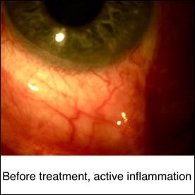
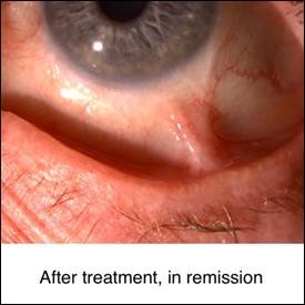
The primary goal of treatment is to suppress active inflammation and decrease the risk of complications. If the cause of uveitis is an infection, appropriate therapy for that infection should be employed with or without anti-inflammatory therapy. Currently, despite recent and ongoing clinical trials, the non-infectious uveitis treatment is based on expert opinion and algorithms proposed by multi-disciplinary panels. A multi-disciplinary approach between pediatric rheumatologists and uveitis specialists is the key for successful treatment. Although corticosteroids are still the first line of treatment of acute uveitis, the major long-term goal of therapy is corticosteroid-free remission with corticosteroid-sparing immunomodulatory therapy. A step ladder approach is commonly employed in the treatment of pediatric patients with uveitis. This approach starts with the use of less potent medications and medications with better safety profiles and shifts to more potent medications with more potential side effects in patients with insufficient response. During corticosteroid free immunomodulatory therapy, high risk blood monitoring tests should be performed at regular intervals. These blood tests include complete blood count (CBC), liver function tests (LFTs), BUN and Cr. Patients on immunomodulatory therapy with conventional medications and biologic response modifying agents should avoid vaccination with live viruses. Almost all conventional immunomodulatory therapies are teratogenic and should be used with caution in child-bearing age.


Corticosteroids

The first line of treatment in pediatric anterior uveitis is often topical corticosteroids. Difluprednate 0.05% is the most potent topical corticosteroid; however, prednisolone acetate 1% is the most commonly used topical corticosteroid in uveitis. While topical corticosteroids are preferred over systemic corticosteroid therapy in most cases, systemic (oral and/or intravenous) and local (periocular and/or intraocular) injections can be considered for resistant, intermediate, posterior, and panuveitis. Slow-release intraocular implants such as dexamethasone intravitreal implant which can last up to 6 months and fluocinolone acetonide 0.19 mg which last for 3 years, are available for more severe cases. Cataract and glaucoma are the most common potential side effects of steroids, especially with intravitreal corticosteroids implants. Oral corticosteroids are used in inflammation recalcitrant to local therapy and in bilateral uveitis. Oral prednisone, at a dose of 1-2 mg/kg of body weight, is the most commonly prescribed oral corticosteroid. However, long-term systemic corticosteroid use should be avoided, especially in the pediatric age group, due to serious side effects including growth retardation, weight gain, hyperglycemia, infection, and osteoporosis. For other types of chronic uveitis in pediatric patients, the recommendations from the American College of Rheumatology/Arthritis Foundation for glucocorticoid use in JIA are employed. These include, 1) superiority of prednisolone acetate over difluprednate topical drops, 2) superiority of adding or increasing topical corticosteroids over systemic corticosteroids for controlling active inflammation, 3) superiority of topical corticosteroid over changing or escalating systemic therapy in patients with flare up on systemic therapy, 4) superiority of adding systemic therapy for patients who need 1-2 drops for controlling the inflammation, 5) in children and adolescents with JIA and CAU still requiring 1–2 drops/day of prednisolone acetate 1% (or equivalent) for at least 3 months and on systemic therapy for uveitis control, changing or escalating systemic therapy is conditionally recommended over maintaining current systemic therapy.14


Non-steroidal Anti-inflammatory Drugs (NSAIDs)

Naproxen and tolmentin have the longest history of successful treatment in pediatric uveitis. It has been shown that celecoxib and diflunisal may be useful in controlling anterior uveitis associated with seronegative spondyloarthropathies. These are not typically the drug of choice for different types of vasculitis.


Conventional Immunomodulatory Therapy

Since long-term use of corticosteroid can incur serious side effects, corticosteroid-sparing immunomodulatory therapy should be the next step in the stepladder approach for the treatment of pediatric uveitis. Conventional immunomodulatory therapies include methotrexate and less commonly used mycophenolate mofetil, azathioprine and cyclosporine. Methotrexate, mycophenolate mofetil, and azathioprine are all anti-metabolites while cyclosporine is considered a T-cell inhibitor. Anti-metabolites and T-cell inhibitors have additive effects on each other; however, anti-metabolites should not be used together preferably.


Methotrexate

Methotrexate is the most frequently used steroid-sparing agent in pediatric uveitis. Methotrexate is considered the first line of therapy in most cases of chronic non-infectious uveitis. The recommended starting dose is 10-15 mg/m2 once a week orally or subcutaneously. Maximum dose is 30 mg/m2; however, doses higher than 15 mg/m2 should be used subcutaneously due to the limited oral bioavailability of methotrexate. A meta-analysis demonstrated that methotrexate was effective in controlling pediatric non-infectious uveitis and the proportion of responding subjects was 0.73 in nine eligible studies. Common side effects of methotrexate include liver, renal and gastrointestinal toxicity. This medication should be used with folic acid supplements to help with normal cell proliferation and prevent side effects.


Mycophenolate Mofetil (MMF)

The optimal dose of MMF for uveitis is unknown; however, the therapy usually starts at a dose of 600 mg/m2 twice a day and the max dose is 3 g/day in two divided doses. There are two reports of MMF therapy in pediatric patients. Although MMF is helpful in the treatment of non-infectious uveitis, it is less effective for arthritis in comparison with other agents. Leukopenia, gastrointestinal discomfort, and hair loss are major side effects.


Azathioprine

Azathioprine can be moderately effective in the pediatric population; however, it is less commonly used due to the increased risk of gastrointestinal side effects in comparison to other anti-metabolites. There are few case series which report the experience with azathioprine in JIA associated uveitis. An observational multi-center study showed that azathioprine may be helpful as a systemic monotherapy or in combination with other immunosuppressive agents in JIA associated uveitis. The safe and well-tolerated dose in children is 3 mg/kg in a single dose or divided dose.


Cyclosporine

It is important to note that cyclosporine is minimally effective as monotherapy; however, the efficacy of cyclosporine increases when it is combined with other immunomodulatory agents. Pediatric dosage of cyclosporine is 2.5 to 5.0 mg/kg/day in divided doses to prevent drug toxicity. Nephrotoxicity, hypertension, hepatotoxicity, hypercholesterolemia, and hirsutism are potential side effects. The dosage of cyclosporine is adjusted based on the medication level. Cyclosporine is infrequently employed in the treatment of uveitis in children.


Biologic Response modifiers

Biologic response modifiers are the next step of the stepladder approach after conventional immunomodulatory agents. These agents block target molecules in the immune system that play a role in the ocular inflammatory process. Therapeutic options include anti-tumor necrosis factor alpha (TNF-α), anti-interleukin 1 (IL-1), anti-B-cell, and anti-T-cell inhibitors. Viral hepatitis and tuberculosis should be ruled out before starting these agents. Out of all the biologic response modifier agents, anti-TNF-α agents have been studied most frequently.


Tumor Necrosis Factor Alpha Inhibitors (TNF-α inhibitors)


Infliximab

Infliximab is a chimeric (human-murine) monoclonal antibody that binds and inhibits circulating and membrane bound TNF-α. The efficacy and safety of infliximab have been studied in retrospective and prospective studies. Infliximab is administrated parenterally with a loading dose of 3-5 mg/kg at 0-2-6 weeks, followed by a maintenance dose of 3-10 mg/kg every 4 weeks. Low dose methotrexate is recommended to prevent antibody formation against the murine part of infliximab molecule. In refractory non-infectious uveitis, a higher dose of 20 mg/kg has been used successfully. Major side effects include susceptibility to infections, reactivation of tuberculosis and histoplasmosis, malignancy, and the development of lupus-like syndrome. Mild to moderate infusion reactions can be seen in 17% of patients.


Adalimumab

Adalimumab, a fully humanized monoclonal antibody, is the only biologic response modifier agent that has been approved by the U.S. Food and Drug Administration (FDA) for the treatment of non-infectious uveitis. Several studies have supported the efficacy of adalimumab in the treatment of pediatric uveitis. Antibody development against adalimumab has been reported in previous studies (13%). This is a favorite medication for ocular immunologists in the treatment of pediatric uveitis. Adalimumab is administered subcutaneously at a dose of 24 mg/m2 up to maximum dose of 40 mg every 2 weeks; however, this interval can be shortened to weekly when the response is inadequate. It was demonstrated in a multicenter, double blind, randomized, placebo-controlled trial (SYCAMORE Trial) that adalimumab therapy-controlled inflammation and was associated with a lower rate of treatment failure than placebo among children and adolescents with active JIA-associated uveitis on a stable dose of methotrexate.

Adalimumab was also shown to be effective in cases of early onset, chronic anterior uveitis with inadequate response to topical therapy and methotrexate.

A meta-analysis performed by Maccora et al. showed the efficacy of adalimumab and infliximab in the treatment of chronic childhood uveitis. However, the results from adalimumab use were superior to those associated with infliximab use in this clinical setting.

The side effect profile of adalimumab is similar to that of infliximab. The high efficacy in pediatric uveitis and ease of administration subcutaneously at home make it a good choice for both the patient and the family.


Abatacept, a T-cell inhibitor biologic agent

Abatacept is a soluble fusion protein which binds to CD80/CD86 on antigen presenting cells and prevents T-cell activation. A recent study described abatacept efficacy in 3 children with idiopathic uveitis. In a study by Tappenier and colleagues, 48% of patients on abatacept failed treatment and it was concluded that abatacept was not a successful treatment in pediatric uveitis. Dosing is 10 mg/kg at 0 and 15 days followed by monthly infusion. Alternatively, abatacept can also be employed as a subcutaneous injection at a dose of 125 mg weekly. Side effects include infection susceptibility, gastrointestinal symptoms, and low risk of malignancy. The efficacy of abatacept in pediatric uveitis is inconclusive.


Tocilizumab, an interleukin-6 (IL-6) inhibitor

Tocilizumab is a humanized recombinant anti-IL-6 receptor antibody which can inhibit T-cell activation, immunoglobulin secretion, and angiogenesis. We recently studied tocilizumab on 8 patients (14 eyes) with refractory JIA associated uveitis. The average duration of follow-up period after starting treatment was 28.6 ± 24.6 months (9-70). Intravenous tocilizumab infusions induced and maintained remission in 5 patients (8 eyes). Vasculitis was resolved within 8 months in all but one patient. We found that intravenous tocilizumab infusion can be an effective and safe method of treatment to induce and maintain remission in resistant JIA associated uveitis. Ramanan et al. studied tocilizumab in patients with anti-TNF refractory juvenile idiopathic arthritis associated uveitis (APTITUDE trial). The aim of this phase two trial was to study the safety and efficacy of tocilizumab in children with juvenile idiopathic arthritis-associated uveitis refractory to both methotrexate and TNF inhibitors; however, they did not meet the primary endpoint of their phase 2 trial to support a phase 3 trial of tocilizumab in patients with JIA-associated uveitis. Currently, there are two ongoing trials to study the efficacy of tocilizumab in noninfectious intermediate, posterior and panuveitis: (STOP-Uveitis) and JIA-associated uveitis (JIA-U). (www.clinicaltrials.gov) Tocilizumab has been proposed to be a promising agent for the treatment of pediatric uveitis; however, more studies are required for the ideal dose and interval. The current dose of medication is 165 mg/week through subcutaneous injections or 8 mg/kg of weight through monthly infusions. Monthly infusions of tocilizumab have been found to be more effective than weekly injections.


Rituximab, a B-cell inhibitor biologic agent

Rituximab is a chimeric antibody that targets the B-cell marker CD20 and subsequently induces B-cell apoptosis. Other mechanisms of action include receptor signaling, calcium flux, complement activation or attraction of antibody-dependent effector cells. Miserocchi and colleagues studied a cohort of 8 patients (15 eyes) with severe refractory JIA uveitis treated with rituximab. The mean number of infusions was 8.75 during mean 47.5 months follow-up (almost one infusion every 5 months). Uveitis was inactive in all patients at the last follow-up visit, and improvement was noted 4 months after the first infusion.

Rituximab is traditionally infused at a dose of 17 mg/kg per infusion which would result in a total dose of 70 mg/kg (4 weekly infusions). The long-lasting effect (6-9 months) of rituximab make it an attractive biologic agent. Rituximab in pediatrics is traditionally given slowly, administered at an initial rate of 0.5 mg/kg/hr for the first hour, and is gradually increased by 0.5 mg/kg/hr every 30 minutes based on the patient’s response (maximum rate of 50 mg/hr).

Side effects include infusion reaction, late neutropenia, hypogammaglobulinemia, heart failure, and rarely lethal progressive multifocal neuroencephalopathy (PMN). IgG monitoring may be useful for patients receiving rituximab since persistent hypogammaglobulinemia that required IgG replacement was noted in 4.2% of patients.


Emerging Biologics

Janus Kinase (JAK) inhibitors have been recently employed in the treatment of patients with uveitis and scleritis in adults. There is an ongoing open-label, active-controlled, safety, and efficacy study of oral baricitinib in patients from 2-18 years old with active JIA-associated uveitis or chronic anterior ANA-positive uveitis.


The recommendation of the American College of Rheumatology/Arthritis Foundation for immunomodulatory therapy in JIA which are mostly applied to other types of chronic uveitis in pediatric group include, 1) subcutaneous methotrexate is superior over oral methotrexate for starting treatment, 2) for a sight-threatening condition a combination of methotrexate and a biologic is recommended for the starting treatment, 3) superiority of other TNF-α inhibitors over etanercept in chronic anterior uveitis (etanercept in ineffective in ocular inflammatory diseases), 4) TNF-α inhibitors dose should be boosted and/or interval should be shortened in inadequate response to standard dose of TNF-α inhibitors, 5) second TNF-α inhibitor should be employed after failing the first one, 6) abatacept and tocilizumab can be employed in patients who fail methotrexate and two TNF-α inhibitors, and 7) all effective medications should be continued for 2 years before tapering therapy.

References

Maleki A, Anesi SD, Look-Why S, Manhapra A, Foster CS. Pediatric uveitis: A comprehensive review. Surv Ophthalmol. 2022;67(2):510-529.


New Recommendations for the Management of Uveitic Glaucoma published

A new landmark publication by leading international experts in uveitis and glaucoma – the Uveitic Glaucoma Interest Group (UGIG). In this publication, the UGIG outlines updated treatment and follow-up recommendations for the management of Uveitic Glaucoma, including a stricter intraocular pressure (IOP) target of 16 mmHg to better protect vision.

Please check out the attached flyer which summarizes these new recommendations on Uveitic Glaucoma! If you’d like to learn more, please visit https://uveiticglaucoma.com/ where you can listen to an AI-generated podcast summary of the article, find a link to the publication, and download the flyer.

Mr J Ronald Quattrocchi

I was treated, very many years ago, at Wills Eye Hospital by (retired since 2021) David Fischer, MD for (now dormant) Uveitis in both eyes. The very first surgical intervention of the right eye (the most effected of the two eyes) facilitated an intraocular implant into the “lens capsule”. A problem occurred in that an opening (or hole) had been surgically made into the original “lens capsule” to permit additional work on retinal damage. The resulting vision provided by these procedures was essentially (for a week or so) edge-to-edge “brightness”. Subsequently, that opening within the lens capsule closed down somewhat and hence resulted in a marked dimming around the edges (bright in the center and shaded all around the edges) of the visual field. Because of this anomaly, the subsequent surgical procedure in the left eye was facilitated wherein the entire lens capsule was removed and the artificial lens was sutured directly into the eye itself.
Albeit somewhat problematic, over the years, I managed my daily activities with respect to the aforementioned problematic right eye surgery. Now, at my advanced age, it appears that my brain is attending (basically and mainly) to vision provided by the (brighter) left eye while occasionally perceiving “visual flashes” of images provided by the somewhat dimmed imagery from the problematic right eye. The resultant vision is distressingly seen as a double image of sorts (as one image provided by the brighter left eye to be interfered, as it were, with angular images provided by the problematic right eye). With respect to my advanced age and the underlying (dormant) Uveitis, can something be done to correct this problem… such as laser treatment to open up the closed-down right eye lens capsule or the complete removal of that eye’s lens capsule and the subsequent suturing (as in the brighter left eye) of the artificial lens implant into the eye itself?

PUK with RA

Hi there, I developed PUK, I have Rheumatoid arthritis since 15 years and I am just on Hydroxychloroquine and NSAIDs for my RA. In the last 2 years my dry eye got worse and I reached the end stage what opthalmology can do for me. Since 2years I fighting with my Rheumatologist to change my disease modifying drugs, I reacted badly to methrotraxate, sulfasalazine ect.
My joints aren’t to bad but all the Inflammation is now in my eyes. Anyone here who was put on any biologicals for PUK with rheumatoid arthritis. My future looks quite bleak and I want to fight for my family, my life. I am a emergency veterinary surgeon and have medical background , sadly there are not many studies out there to proof to my rheumatologist to change my disease modifying drugs. Thanks

Golimumab/Simponi is denied by insurance — what to do?

I’ve been on Simponi for about a year for PIC (after being on Humira and developing antibodies to that). The Simponi is working so far, but my new Aetna medicare advantage insurance plan is denying any refills, after 3 appeals. They say it’s “off label” which is true, but FDA has not approved anything else that works for me. So now I have no medication. Any advice on how to deal with this? Simponi is so expensive out of pocket. Hire a lawyer? Switching out of Medicare Advantage to a traditional Part D plan? Thank you for any advice you can provide.

Am I being over prescribed medication?

I have been diagnosed with birdshot choryoretinopathy and have been prescribed Mycophenolate (500 mg) , one pill twice daily for 2 weeks, then two pills twice daily for another two weeks, until three pills twice daily.
Is that the usual prescription? It seems to be over prescribed. Why not keep to the one pill twice daily? I do not have many other symptoms- other than a retinal membrane and uveitis.

Adalimumab therapy

Hey everyone, I’m new to the forum. Since 2020, I have been dealing with non-infective anterior uveitis HLA B27 positive, managed successfully with Prednisolone eye drops. Due to frequent recurrences this past year, my immunologist is considering adalimumab therapy to reduce Prednisolone usage. I’m seeking experiences with adalimumab biosimilars or information on the risks and benefits of this treatment. Thanks in advance.

Birdshot Retinochoroidopathy

Birdshot Retinochoroidopathy

C. Stephen Foster, MD, FACS, FACR

What is Birdshot Retinochoroidopathy?

Birdshot retinochoroidopathy, commonly referred to simply as “birdshot”, is a rare form of posterior uveitis which mainly affects the retina and choroid. The disease occurs in women more often than men, typically Caucasian, and most often between the ages of 30 and 60. “Birdshot” can be a severe and blinding disease if unrecognized or undertreated.

What causes “birdshot”?

The etiology of the disease is, as yet, unknown. There is a class of genes known as human leukocyte antigens (HLA), and specifically one called HLA-A29 (also HLA-A29.2 or HLA-A*2902), that is present in the overwhelming majority of “birdshot” patients, and is thought to be involved in the development of the disease. Infection, by virus or bacteria, in susceptible individuals is thought to act as a trigger, with the disease then being self-propagated by an autoimmune mechanism.

What are the symptoms of “birdshot”?

It is normally a chronic problem, with symptoms that develop and progressively worsen over several months to years. The most common complaints are:

  • Floaters and flashes
  • Blurry or hazy vision, sometimes described as looking through murky water
  • Decreased color and/or night vision

Patients usually do not complain of pain or redness, though it can be present. There are not any systemic diseases that are typically associated with “birdshot”.

How do you diagnose “birdshot”?

Clinical examination is the most important tool in the diagnosis of “birdshot”, though a full history and review of systems must be performed to help evaluate for other potential infectious or autoimmune causes. Examination often shows little to no inflammation in the “front” of the eye, however dilated exam can reveal a dense collection of inflammatory cells and debris in the vitreous, pallor of the optic nerve, attenuation of retinal vessels, retinal vasculitis, macular edema, and the presence of “birdshot lesions” or creamy yellow-white spots involving the retina and choroid. Cataract formation can also occur as a result of long-standing inflammation or chronic use of corticosteroid eye drops. Rarely, new blood vessels can sometimes grow between choroid and retina which can cause severe vision loss even after inflammation is treated.

What testing is performed to help diagnose “birdshot”?

A large array of clinical testing can be very helpful, starting with checking blood work, especially for the presence of the HLA-A29 gene when “birdshot” is suspected. Color vision testing can show mild to severe deficit. Fundus autofluorescence may reveal abnormal areas of hypopigmentation that correspond to “birdshot lesions” or overlap areas or normal appearing retina. Fluorescein angiography may show window defects in areas of “birdshot lesions” or evidence of macular edema, retinal vasculitis, optic nerve inflammation, or choroidal neovascularization. Indocyanine green (ICG) angiography is used to evaluate the choroid, and can show hypolucent lesions that may not be visible on dilated exam. Visual field testing with a blue-on-yellow protocol (shortwave automated perimetry, or SWAP) is more sensitive than normal white-on-white visual field testing, and may show more advanced field defects. Optical coherence tomography (OCT) can reveal macular edema or epiretinal membranes. Lastly, and perhaps most importantly, standard electroretinogram (ERG) can be a very useful tool in both the diagnosis of “birdshot”, and in assessing response to therapy, particularly the 30-Hertz photopic protocol, which will often show delayed implicit times and decreased signal amplitudes that may improve with treatment.

Can “birdshot” be treated?

Yes, this disease is very treatable, and after proper treatment, it is even possible to achieve long-term remission of active inflammation while off all medications. Unfortunately, damage done to retina and choroid, and to most other parts of the eye affected, is irreversible, which makes timely recognition and initiation of therapy the key to a good outcome and preserving vision.

How do you treat inflammation from “birdshot”?

“Birdshot” is a severe and stubborn form of uveitis, and as with all types of uveitis, inflammation must be quieted by whatever means necessary or blindness will ensue.

Corticosteroids
Coritosteroids are usually the way to treat most non-infectious uveitis quickly, but for “birdshot”, the way in which they are given may have an effect on the disease course. Our research at MERSI has found that patients who have received oral corticosteroids have shown a higher rate of disease recurrence, even after achieving long-term remission on immunomodulatory therapy. Injection into the eye (intravitreal injection) was not shown to have this risk. Topical eye drops are not effective in treating inflammation in the back of the eye.

Immunomodulatory Therapy
Immunomodulatory therapyis the standard of care for treating birdshot retinochoroidopathy. Our years of experience at MERSI treating patients with “birdshot” have shown the most effective initial treatment strategy involves starting combination therapy with mycophenolate mofetil (CellCept®), an antimetabolite, and modified cyclosporine A, a calcineurin-inhibitor. In cases of intolerance or poor efficacy, mycophenolate is sometimes replaced with another antimetabolite, or the patient is transitioned to intravenous therapy with a TNF-α inhibitor such as infliximab (Remicade®). Stubborn and poorly controlled disease on these medications may require use of an alkylating agent such as cyclophosphamide (Cytoxan®).

Corticosteroid Implant
A corticosteroid implant surgically placed within the eye, fluocinolone (Retisert®), can be used for patients who are not able to achieve remission on or tolerate immunodulatory therapy, or in cases where this option presents less of a burden on life than long-term medical therapy.

What other problems are associated with “birdshot” and how are they treated?

Cataracts and Glaucoma
Cataracts and glaucoma can both occur as a result of active inflammation or long-term treatment with corticosteroids.

  • Cataracts can be removed, ideally when inflammation has been treated and the eye is quiet, but may need to be removed to see and treat problems inside the eye.
  • Glaucoma is initially treated with eye drops, followed by laser therapy, and lastly surgery if necessary.


Retina
Problems with the central retina, especially macular edema and epiretinal membranes, are frequently seen.

  • Macular edema may resolve when inflammation is treated, however if it persists, it can also be treated with topical NSAIDs or oral acetazolamide.
  • Epiretinal membranes can be surgically removed if they are felt to cause progressively worsening vision. Intraocular injection of either corticosteroid or a VEG-F inhibitor, such as bevacizumab (Avastin®), is extremely effective in treating macular edema. These can also be used to treat choroidal neovascularization, new blood vessels that grow between retina and choroid as a result of a break in the barrier that lies between them.

Durezol vs immunosuppressant therapy

Originally, I was diagnosed with APMPPE in 2017, and did not have uveitis, just the beginnings of an epiretinal membrane. Unfortunately, I developed uveitis within the past year, and my new Opthamologist has declared my condition to be Birdshot chorioretinopathy (as I now have tested positive for HLA-A29). I am on Durezol, and have been sent to a rheumatologist in order to receive Mycophenolate. Is this a normal procedure? Which medication works best for Birdshot/APMPPE (with fewer side effects)?

Resources for those not getting adequate treatment

Hi, I’m 26F and have been dealing with intermediate uveitis since May 2023. I’ve had a trickle down of neurological symptoms since March 2023, new/worsening symptoms over time are severely affecting me and my life. I was on a low dose of prednisone up until October 2023, which helped subside the eye inflammation but did not clear it and I experienced new eye symptoms while I was on it. I am still having disruptive floaters/snow vision, fireworks/glittering, blur/haze, sometimes sharp pain and double vision.

Lumbar Puncture in July 2023 with lymphocytic pleocytosis, elevated CSF igG index, CSF Oligoclonal bands. Repeat LP in October 2023 revealed 6x higher inflammation, negative for infectious viral/bacterial/fungal causes. MRI with small non-specific non-enhancing bilateral subcortical hyperintensities in frontal lobes as of August 2023. Clear thoracic/cervical spine MRI as of August 2023. No blood inflammation or abnormalities other than elevated beta2glycoprotein antibodies. No abnormalities on chest/abdomen/pelvis CT. No previous medical conditions, events, surgeries or hospitalizations.

I have not had any testing on my eyes other than photography.

Doctors are currently unwilling to provide further treatment for the inflammatory immune response that I am having because they don’t know the ‘exact cause’ and because my findings/symptoms do not fit clearly inside of a diagnostic box. I am just getting worse and my symptoms are not improving at all. I’m not able to live a normal life and don’t know what to do/where to go from here – I have sought multiple opinions from various specialists. I am on state Medicaid insurance, so I cannot afford to see anyone outside of my state.

Are there any resources available for this type of situation or universities/clinics that would be willing to evaluate me as a patient outside of my state (VA) that would be more knowledgeable/better equipped to deal with a complex case?

Answer: Hi, so sorry to hear your story of obstacles to care.  I think this is more of a clinical question and needs to some clarification on your history so I would ask that you provide your email so we can communicate.  Please use the conus link on uveitis.org and email the support group link.  Thank you.  Frances Foster NP

ESSENTIAL Medical Visits requiring an Office Appointment

COVID19 Patient Information Update for MERSI Eye Center and OIUF

As you may have heard, Governor Baker has issued “A Stay in Place Order” for the state of Massachusetts for all non-essential businesses beginning at noon on March 24, 2020.   MERSI is considered essential, but we will need to restrict visits to those defined as essential.

ESSENTIAL Medical Visits requiring an Office Appointment:

  • Any patient actively being treated with immunosuppressive therapy (oral, infusion, or injection)
  • Any patient with acute or new symptoms/problems
  • Any follow up patient who had acute or new symptoms/problems and was placed on active treatment needing reassessment
  • Patients who are experiencing flashers, floaters, eye pain, or light sensitivity
  • Retina patients who are receiving injections to control their eye disease progression
  • Glaucoma patients actively being treated for elevated pressure issues or in late-stage disease
  • If you  feel the situation is urgent, call to schedule an appointment or arrange for a phone call with one of our clinical staff to determine the nature of the urgency for the visit
  • All routine, annual, screening appointments will be rescheduled unless you have new symptoms (which can be discussed with one of our clinical staff to determine urgency)

****We ask if you are sick or have returned from travel or been exposed to someone from who had traveled or is high-risk for contracting the Covid 19 virus to reschedule your appointments.***

Recommendations for those of you on immunomodulatory therapy:

We recommend you always  be vigilant about practicing universal precautions to prevent getting ill (see below).  The flu is a concern each year, which is why we recommend and encourage you to get the flu shot and now, we have a new viral illness in addition to the flu that we all are at risk for contracting.

Coronavirus disease (COVID-19) is an infectious disease caused by a new virus that had not been previously identified in humans.

The virus causes respiratory illness (like the flu) with symptoms such as a cough, fever and in more severe cases, pneumonia.

HOW IT SPREADS:

The new coronavirus like the flu spreads primarily through contact with an infected person when they cough or sneeze, or through droplets of saliva or discharge from the nose.

Practices to Protect Yourself:

  1. We encourage universal precautions, which one should practice always to avoid the flu, cold, or other illnesses in addition to the Coronavirus.
    1. 20-30 second handwashing, use hand sanitizer if no soap after touching surfaces and always before eating
    2. disinfect surfaces you will need to touch including your work space
    3. avoid touching door knobs or handles, elevator buttons, shopping carts, etc with hands. Use instead for example a wipe, paper towel, shirt sleeve, glove, or on elevator button your knuckle instead of finger tip  
    4. do not eat food with hands  
    5. avoid touching your face, mouth, and nose with hands
  • Air Travel or public spaces Postpone to avoid any risks until the virus spread is under control. 
  • The CDC provides great guidelines on their website. Please visit www.cdc.gov

Of note, If you develop any illness, infection, or fever of 99 degrees F or > or if you have been exposed to a high risk person for Covid 19 or Flu; please notify your doctor immediately and hold your immunosuppressive therapy medication immediately.  Specifically, if you have a fever of 101° Fahrenheit (38.3° Celsius), with or without chills, call your doctor immediately.   If you cannot reach your doctor, go to an emergency room.

In general, when you are healthy your risk for contracting an illness lessens so we recommend 8 hours of sleep, good hydration (8-10 glasses of noncaffeinated fluid a day), a multivitamin daily, and exercise if possible 3 times a week.   We also recommend smoking cessation if applicable to you.

Mersi Providers and Nursing Staff


Recent Research Relating to Cicatricial Pemphigoid and the Use of Intravenous Immunoglobulin (IVIG)

C. Stephen Foster, MD, FACS, FACR

Dr. Foster et al. recently published a chart review of all Ocular Cicatricial Pemphigoid (OCP) patients seen at the Massachusetts Eye Research and Surgery Institution (MERSI) between 2005 and 2015 to look at the management of OCP with Intravenous Immunoglobulin (IVIG) as the only therapy. IVIG infusion was administered in the usual manner with the dose being two grams per KG of weight and the total dose divided for infusion over three consecutive days every month.

Five hundred and twelve (512) patients were identified with OCP at MERSI, and only17 patients or 34 eyes were treated with IVIG as the only therapy.  Seven were female, and ten were male with the average age at diagnosis as 60.7 years old.  The follow-up time ranged from 12 to 140 months. Twenty-six eyes (76.5%) achieved remission. Nine remission eyes received cataract surgeries, and 2 of them had a relapse (22.2%). The other 17 eyes did not undergo ocular surgery and remained in remission.

The findings revealed that IVIG as only therapy showed high efficacy in stage one on Foster staging scale of OCP (7/7, 100%).  Ocular surgery can be associated with OCP relapse.  The conclusion from the chart review study was IVIG monotherapy is an effective and safe therapy in patients with stubborn OCP.  However, ocular surgery can be associated with OCP relapse even when a patient was in remission.  Rituxan and IVIG combination are still top of the line treatment for OCP.

#ocularcicatricialpemphigoid #ocp #intravenousimmunoglobulin #IVIG

Cost of Care of Patients with Uveitis

C. Stephen Foster MD

Increasingly restrictive “gate keeping” policies of health maintenance organizations, insurance companies, and other medical insurance plans have created increasing pressure on ophthalmologists to be parsimonious in their use of medical services in both the diagnostic and therapeutic care of patients with a variety of medical disorders, including those with uveitis. These pressures are particularly prominent in the physician’s care of patients with chronic disorders, and ophthalmologists caring for patients with uveitis are increasingly experiencing this restrictive pressure. We wondered what the cost of diagnostic and therapeutic care of a patient with uveitis might be, given what we, as a uveitis referral center, see as appropriate yet fiscally prudent care. The cost of care obviously varies greatly, depending on the underlying cause and on the severity of the patient’s uveitis and associated complications. As a first step in estimating the total annual direct cost in the care of patients with uveitis we restricted our analysis to patients with HLA-B27 associated uveitis. We also restricted our analysis to the direct medical cost of caring for such patients, recognizing that direct non-medical costs, indirect morbidity costs, and other intangible economic loss costs, disability payments, absences from work, etc. are real but difficult to measure costs of the total cost of the patient’s illness. Direct medical costs are transactions and expenditures for medical products and services, including diagnostic studies, physician fees, hospitalization costs, surgical costs, rehabilitation and subsequent long-term care costs.

A cohort of 105 patients with HLA-B27 associated uveitis were studied in 2002. The diagnosis in each instance was established on our Service, and a minimum follow-up of two years existed for each patient. The medical records were reviewed for the diagnostic studies and costs of each performed, the physician and hospital fees associated with visits and/or surgery, in the cost of medical therapy. The average direct annual cost per patient per year was calculated. A stepladder approach to therapy was employed in an effort to eliminate recurrences of uveitis. The first step on the stepladder was the use of steroids, through any route required to achieve the goal of quieting the uveitis. Oral non-steroidal anti-inflammatory agents were added, if recurrence typically continued despite the use of steroids. Immunosuppressive chemotherapy was employed if patients continued to have recurrence of inflammation despite the use oral non-steroidals. Ten patients eventually required the use long-term oral immunosuppressive agents, and 30 patients were on chronic oral non-steroidal anti-inflammatory drugs. The average annual cost of care of the patients with HLA-B27 associated uveitis was $4,108.60 (range $433 to $9,683.18). These results reflect an average cost of caring for a cohort of patients with recurrent HLA-B27 associated uveitis of varying severity. The results may serve as an indicator, to health maintenance organizations and other pooled-risk insurers, of the cost of prudent care of patients with this form of uveitis. We would emphasize that we were very cautious and parsimonious in our use of laboratory tests and frequency of return visits, striving for the greatest degree of economy, while at the same time striving for the best possible outcomes (for outcomes analysis studies performed on these and other patients with uveitis, please refer to the Bibliography section of this Web Site). Clearly, patients with recurrent or chronic uveitis require significant expenditure of the health care dollar. It is, however, money well spent, since the preservation of sight from modern care of such patients profoundly reduces the prevalence of blindness secondary to uveitis, and hence reduces the economic burden on our society in total.

Did you know the Connection between Arthritis and Ocular Disease?

Connection Between Arthritis and Ocular Disease
C. Stephen Foster, M.D.

The eye is made up primarily of collagen, as are ligaments, tendons, and tissue within joint spaces. It is, perhaps, primarily because of this similarity in composition that the eye is often affected by many of the same diseases which affect joints. Some of these disorders include Juvenile Rheumatoid Arthritis, Adult Rheumatoid Arthritis, Systemic Lupus Erythematosus, Relapsing Polycondritis, Behcet’s Disease, Granulomatosis with Polyangiitis (formerly called Wegener’s), Polyarteritis Nodosa, and Scleroderma or systemic sclerosis. Additionally, the type of vasculature that is present in the eye has special characteristics that produce an extraordinarily sensitive “barometer” or “sentinel canary” in the eye for potentially lethal vasculitis that can be associated with the aforementioned collagen vascular diseases. Specifically, we know from considerable experience that, despite the fact that a patient’s rheumatoid arthritis may be “burned out” as far as active inflammation of the joints in concerned, nonetheless, the patient may well have subclinical rheumatoid vasculitis affecting various internal organ systems. The eye is a very potent indicator of such subclinical potentially lethal vasculitis, and if the eye becomes involved with retinal vasculitis, uveitis, scleritis, or peripheral ulcerative keratitis in such a patient, we take that as a very strong signal that the patient must be evaluated extremely carefully for potentially underlying vasculitis affecting viscera and we also take such a potentially blinding ocular lesion very seriously from the standpoint of the need for aggressive systemic immunomodulatory therapy in order to prevent permanent damage to the eye from such lesions.

For example, we have seen many instances in which patients with systemic lupus erythematosus appear, systemically, to be doing quite well (indeed, the patient’s Rheumatologist has told her that she is doing very well) despite the fact that new-onset uveitis, scleritis, or retinal vasculitis has developed in one eye. We have seen this story evolve to life-threatening central nervous system vasculitis and/or lupus renal disease when the onset of the ocular inflammation was not taken as an indication for increasing the vigor of systemic therapy. We have tried diligently, therefore, over the past 15 years to raise the consciousness, not only of ophthalmologists worldwide, but also of rheumatologists and other internists of the valuable indicator that the eye can be with respect to seriousness of associated arthritic/collagen vascular disease.

Pediatric Uveitis

Uveitis is the third leading cause of blindness in America, and 5% to 10% of the cases occur in children under the age of 16. But Uveitis in children blinds a larger percentage of those affected than in adults, since 40% of the cases occurring in children are posterior uveitis, compared to the 20% of posterior Uveitic cases in the adult Uveitis population.

There are, at any one time, approximately 115,000 cases of Pediatric Uveitis in the United States, with 2,250 new cases occurring each year. Spread across the entire U.S. population, therefore, and across all offices of Ophthalmic practitioners, the likelihood that any one individual practitioner will care for a patient with Pediatric Uveitis is relatively small, and the likelihood that any single individual will have significant experience in caring for large numbers of cases over a long period of time is vanishingly small. This accounts, we believe, at least in part for the sub-optimal care that many of our children with Uveitis appear to be receiving, even in these “modern” times. The stakes are incredibly high, for the child, for the parents who will be faced with (usually) many years of dealing with this health problem in their child, and for society at large because of the life-time of dependence which occurs in those who eventually reap substantial visual handicap as the result of sub-optimal treatment.

We believe that current epidemiologic data emphasize two critically important goals for all of us in Ophthalmology, acting together, in an effort to change the current prevalence of blindness caused by Pediatric Uveitis:

  1. Repeatedly emphasizing to parents, other medical colleagues, especially Pediatricians, and school personnel the critical importance of routine (annual) vision screening for all children.
  2. The critical importance of beating back the frontiers of general ignorance and mind sets, eliminating the all-too-common pronouncement by physicians to parents of a child with Pediatric Uveitis that:
  3. “He’ll (She’ll) out grow it.”
  4. “The drops will get him (her) through it.”
  5. “It’s just the eye; systemic therapy is not warranted.”

Statements (a) and (b) are true, but too often pull the doctor, and patient, and family into the seduction of nearly endless amounts of topical steroid therapy. It is generally true that the child will in fact “out grow” the Uveitis, i.e., that the Uveitis will no longer be a problem eventually. The pity is, however, that so often by the time the child “out grows it”, permanent structural damage to retina, optic nerve, or aqueous outflow pathways has already occurred, and the blinding consequences are now permanent. It is also true that for any individual episode of Uveitis, the steroid drops usually will get the patient through it. But the fact is that so many children with Pediatric Uveitis have recurrent episodes of Uveitis such that the cumulative damage caused by each episode of Uveitis and the steroid therapy for each episode eventually produces vision-robbing damage. And item (c) is simply the result of the common myopic viewpoint of Ophthalmologists: that it is just an eye problem, and therefore should simply be treated with eye medications. Nothing could be further from the truth! And unless and until large numbers of Ophthalmologists reframe this socially and epidemiologically important matter, the prevalence of blindness secondary to Pediatric Uveitis is not going to change.

The differential diagnosis of Pediatric Uveitis is relatively vast, and therefore the detective work required to properly pursue the underlying diagnosis is complex. The job can be slightly simplified by “playing the odds”, categorizing the case as carefully as possible into anterior non-granulomatous; anterior granulomatous; intermediate; posterior, with vasculitis; posterior, without vasculitis; and categorizing it into the general age groups of Infancy (0 to 2 years), Toddler-School Age (2 to 10 years), and Adolescence (10 to 20 years).

The most common etiologic groups in children segregated into these groups are shown in Tables 1-6

TABLE 1 (Anterior Non-Granulomatous)

Idiopathic

HLA-B27 associated

Juvenile Rheumatoid Arthritis

Ankylosing Spondylitis

Reactive Arthritis (formerly called Reiter’s syndrome) disease

Psoriasis

Inflammatory bowel disease

Nephritis

Systemic lupus erythematosus

Herpes Simplex virus

Lyme disease

Leukemia

Drug-induced

TABLE 2 (Anterior Granulomatous Uveitis)

Sarcoidosis

Inflammatory bowel disease

Syphilis

Herpes simplex virus

Tuberculosis

Bechet’s disease

Multiple Sclerosis

Fungal disease

Whipple’s disease

Leprosy

TABLE 3 (Intermediate Uveitis)

JRA

Pars Planitis

Multiple Sclerosis

Lyme disease

Sarcoidosis

TABLE 4 (Posterior Uveitis, without vasculitis)

Toxocariasis

Toxoplasmosis

Leukemia

Tuberculosis

Intraocular Foreign Body

Vogt-Koyanagi Harada Syndrome

TABLE 5 (POSTERIOR UVEITIS, with vasculitis)

Posterior Uveitis with vasculitis

Cytomegalovirus

HSV/VZV

Inflammatory bowel disease

Syphilis

Bechet’s disease

Systemic lupus erythematosus

Kowasaki’s disease

Sarcoidosis

Polyarteritis nodosa

Granulomatosis with Polyangiitis (formerly called Wegener’s)

TABLE 6 (most common causes of Uveitis in infants)

Herpes Simplex Virus

Toxocara

Congenital Loes

Retinoblastoma

TABLE 7 (most common causes of Uveitis in Toddlers/School Children)

Toxocariasis

Toxoplasmosis

Leukemia

Vogt-Koyanagi Harada Syndrome

Diffuse Unilateral Sclerosing Neuroretinitis

Juvenile Rheumatoid Arthritis

TABLE 8 (most common causes in Adolescents)

JRA

Pars Planitis

VKH

Toxoplasmosis

HLA-B27-associated sarcoidosis

Bechet’s disease

Intraocular Foreign Body

We believe that aggressive efforts should be made to uncover the underlying cause of Uveitis in any child. If the review of systems is negative and the patient has non-recurrent anterior granulomatous Uveitis, we would not do laboratory studies. However, if review of systems is positive, we would “follow the review of systems”.

For recurrent anterior non-granulomatous Uveitis we would obtain a complete blood count and urine analysis, ANA testing, HLA-B27 testing, and would “follow the review of systems”.

The diagnostic step ladder in a pediatric patient with anterior granulomatous Uveitis, recurrent or not, would include a CBC with urine analysis, and FTA-ABS testing, Lyme disease antibody and western block, PPD analysis, chest X-ray, ANA, and angiotensin converting enzyme determination. Chest CT, and Gallium scanning would be pursued if diagnosis of sarcoidosis was strongly suspected, and, of course as usual, we would “follow the review of systems positives”.

In a patient with intermediate Uveitis, all would deserve laboratory evaluation, including CBC, urine analysis, chest X-ray, FTA-ABS, ACE, PPD, Lyme, and ANA titers.

Any patients with posterior Uveitis would deserve an extensive vasculitis work-up, if vasculitis were present, and a search for “the usual suspects” with an eye to an infectious etiology, such as that producing a granuloma in the choroid in a patient with toxocariasis or Toxoplasmosis. An audiogram or lumbar puncture would be done if positive on the review of systems were found such as tinnitus and/or meningeal signs or symptoms. Finally, a diagnostic vitrectomy would be added to the step ladder in a patient with posterior Uveitis if all non-invasive studies were unrevealing and the case was difficult to treat successfully.

On the matter of treatment, here too we believe strongly in the step ladder approach, always beginning with steroids, in any route required to achieve the desired goal, i.e., abolition of all active inflammation. Topical steroids would be followed by an examination under anesthesia with regional steroid injection therapy in a patient with granulomatous or non-granulomatous anterior Uveitis. Systemic steroids would be employed in the event that this approach did not achieve the goal of abolition of all active cells. We are extremely reluctant to get involved with long term daily systemic steroid use in a youngster, because of the obvious growth-retarding properties of such therapy. But long term oral non-steroid anti-inflammatory drug therapy, managed by a Pediatrician, can be extremely successful, in our experience in approximately 70% of children with recurrent non-granulomatous anterior Uveitis. If this strategy is not successful, then consideration of once weekly, low-dose, Methotrexate or daily Cyclosporine or CellCept would be the next considerations.

In granulomatous disease topical steroids often are not sufficient, and systemic therapy, particularly with oral non-steroidal inflammatory drugs, may be utilized sooner rather than later.

With Intermediate Uveitis topical steroids are not effective in penetrating to the level of inflammatory focus. Regional steroid injections or systemic steroids are employed to treat that area, sometimes with adjunctive topical steroids for anterior chamber “spill over” reaction. Retinal Cyropexy can be effective in selected cases of recurrent Pars Planitis, as can therapeutic Pars Plana Vitrectomy. Systemic immunomodulatory therapy, as usual, represents the final step in the step ladder approach in the aggressiveness of care.

Patients with Posterior Uveitis of course do not respond to topical therapy and therefore require systemic steroids and/or immunomodulators right from the very beginning.

We hope that this will provide some help to those Ophthalmologists who have also concluded that the usual approach to Pediatric Uveitis, i.e., steroid drops, is not always sufficient, but who are hesitant to take the initiative to commit the patient to more aggressive treatment.